What Is a Dental X-Ray Sensor?
A comprehensive clinical guide explaining digital intraoral x-ray imaging technology, mechanical principles, and application parameters of a modern dental x-ray sensor.
⏱️ 12-15 min read
✓ Clinical Review Complete
⚡ Quick Answer Summary
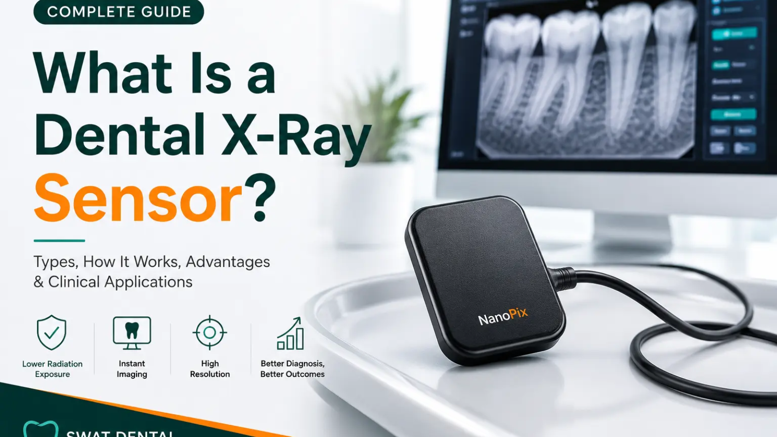
A dental x-ray sensor is an electronic diagnostic device placed intraorally to capture high-definition radiographic pictures of oral structures. Replacing traditional plastic films, these modern units utilize advanced solid-state semiconductors (specifically CCD or CMOS sensors) to convert raw radiation streams instantaneously into a digital display format on a computer screen. By utilizing a high-efficiency dental x-ray sensor, clinics eliminate physical chemical developers, decrease patient radiation by 50% to 80%, and yield immediate, accurate diagnostics.
Table of Contents
1. What is a Dental X-Ray Sensor?
A digital dental x-ray sensor is an extremely precise intraoral medical screening asset crafted to isolate, map, and document the sub-surface physiological structures of a patient’s mouth. These sensors represent a quantum leap from traditional silver-halide film canisters, introducing an electronic silicon grid interface that acts dynamically upon exposure. When implementing a dedicated dental x-ray sensor configuration, it fits comfortably behind dental arches to convert invisible electromagnetic rays directly into a stream of structured numerical visual arrays.
The global elimination of physical development chemical baths introduces safer, instantaneous diagnostic environments to operatory teams. By using a top-tier dental x-ray sensor, operators can effortlessly review crystal-clear bone structural patterns, underlying abscess tracks, and micro-cavities without any standard processing lag.
Key Characteristics of a Dental X-Ray Sensor:
- Digital Capture Engine: Transforms energetic raw particle impacts straight into clean pixel data matrices.
- Real-time Display Mechanics: Streamlines imaging output onto computer terminal screens within milliseconds of trigger fire.
- Drastic Dosage Minimization: Operates at peak diagnostic efficiency with up to 80% lower dose exposure rates than heritage film.
- Ergonomic Forms: Standard dental x-ray sensor profiles are configured with rounded corners and optimized shapes to limit soft-tissue patient discomfort.
- Dynamic Image Tuning: Enables doctors to magnify, adjust contrast, alter color filters, and accurately trace density levels using diagnostic software suites.
2. Types of Dental X-Ray Sensors
Modern operatory layouts generally implement one of three distinct diagnostic device categories depending on workflow requirements, budget, and mobility setups. Selecting the ideal dental x-ray sensor architecture depends heavily on your clinic’s imaging demands:
CCD (Charge-Coupled Device) Technology
Technology Principle: CCD systems implement a dedicated array framework built on a foundational layer of specialized crystalline silicon. When radiation streams meet the chip face, pixel nodes pass their localized charges sequentially across the chip row toward a single readout node to create a uniform output signature across the dental x-ray sensor surface.
- Industry-best image uniformity with virtually zero internal signal variance.
- Exceptional low-noise performance parameters for high-definition diagnostic tracking.
- Wired configuration architectures requiring stable peripheral power attachments to the dental x-ray sensor unit.
- Thicker mechanical housing compared to modern thin CMOS profiles.
Best Suited For: Advanced endodontics, clinical maxillo-facial surgery tracks, and institutions emphasizing maximum image depth profiles.
CMOS (Complementary Metal-Oxide Semiconductor) Hardware
Technology Principle: CMOS technology alters tracking frameworks by housing individual transistor and amplifier modules inside every solitary pixel node. Rather than moving charges across rows sequentially, each node on this type of dental x-ray sensor measures, amplifies, and pipes out its unique electrical charge line instantly.
- Extremely fast image acquisition with zero data transmission lag.
- Low energy draw allows for completely wireless Bluetooth or Wi-Fi dental x-ray sensor configurations.
- Thinner profile boundaries, significantly increasing patient comfort inside narrow jaws.
- Highly cost-effective manufacturing pipelines making them globally accessible.
Best Suited For: General high-turnover dental clinics, pediatric operatory environments, and settings emphasizing streamlined wireless ergonomics.
PSP (Photostimulable Phosphor) Plates
Technology Principle: PSP systems act as an elegant middle-ground bridging legacy film handling with modern digital files. These thin, cordless plates contain high-grade phosphor crystals that store structural energy paths upon ionizing contact, serving as an alternative to a rigid dental x-ray sensor. They require post-exposure placement into a laser tracking scanner that reads out and builds the final computer display image.
- Completely identical physical thickness and flexibility compared to classical plastic films.
- Wireless freedom during placement; zero cables extending from the mouth.
- Plates can be reused hundreds of times when handled with proper hygienic barrier jackets.
- Requires a physical scanning station step which introduces a minor 10-25 second operational delay.
Best Suited For: Orthodontic practices tracking tooth migration lines, community outreach missions, and offices migrating gradually from legacy setups.
3. Step-by-Step Operational Engineering Principles
The conversion from a raw radiation burst into an observable digital diagnosis requires precise timing. Each phase across the dental x-ray sensor assembly must integrate flawlessly to ensure artifact-free visual files:
The process initiates at the tubehead assembly, where a specific electrical input (typically calibrated around 60-70 kVp) heats a tungsten filament. This fires a targeted stream of x-ray photons through the localized area of the patient’s face toward the target tooth structures.
As the energetic particle path intersects the jawline, it faces varying absorption properties based entirely on material density. Mineralized enamel and dense jawbone shield and arrest a massive fraction of photons, while soft vascular tissue and air cavities allow particles to sail through toward the active dental x-ray sensor face.
Before interacting directly with the raw silicon layer inside the dental x-ray sensor, the incoming x-ray energy hits an internal layer called a scintillator (commonly cesium iodide). This specialist layer converts invisible x-ray radiation down into a clean, uniform burst of visible light wavelengths.
The newly transformed light photons pass directly into the pixel well structure of the semiconductor. This interaction triggers an internal photoelectric phenomenon, creating localized electrical changes across the dental x-ray sensor array. Dense regions create minimal charge structures, while clear areas fill wells with maximum voltage indicators.
An internal analog-to-digital converter (ADC) systematically counts every localized voltage value on the dental x-ray sensor chip and translates it into standard binary code data. This raw digital packet is instantly routed via high-speed USB paths to server platforms, rendering a crisp, 256-grayscale diagnostic image.
4. Core Technical Advantages
Adopting modernized solid-state electronic imaging technology completely transforms your clinical workflow efficiency while elevating patient safety parameters across the board. Implementing a high-quality dental x-ray sensor provides measurable benefits:
Radiation Protection Maxima
Digital silicon substrates exhibit remarkable sensitivity to incoming light bursts compared to standard physical film coatings. This specialized dental x-ray sensor responsiveness allows operators to reduce patient radiation dosage numbers by up to 80% per diagnostic exposure run.
Instant Diagnostics Integration
Completely cuts out physical tracking, chemicals, fixed darkroom spaces, and processing delay cycles. Diagnostic dental x-ray sensor configurations materialize on target screens under 3 seconds, significantly reducing overall operational chair time.
Unmatched Resolution Detail
Offers unmatched line-pair resolution densities per millimeter. A premium dental x-ray sensor enables clinicians to instantly apply high-power digital magnification matrices, invert gray channels, and pinpoint structural shifts easily.
Streamlined Database Storage
Stores comprehensive data records on local server paths or cloud-backed networks. This safely clears physical storage rooms and guarantees instant image retrieval options whenever duplicate insurance copies are needed.
Long-term Overhead Reduction
Completely eliminates ongoing tracking expenses tied to chemical purchases, developer machine maintenance, and disposal regulations. While the initial dental x-ray sensor investment is higher, it pays off with low per-exposure maintenance costs.
Eco-Friendly Workstations
Removes hazardous lead elements and dangerous toxic fixer fluid compounds from the clinic environment, keeping operations cleanly aligned with modern environmental guidelines.
5. Technical Comparison Matrix
This detailed technology matrix highlights the clear differences across modern digital devices to guide your dental x-ray sensor procurement decisions:
| Performance Attribute | CCD Sensor Systems | CMOS Sensor Systems | PSP Plate Formats |
|---|---|---|---|
| Diagnostic Resolution | Maximum/Ultra-High Linearity | High/Excellent Precision Levels | Very High Resolution Limits |
| Image Generation Velocity | Instantaneous (Under 2 Seconds) | Instantaneous (Under 2 Seconds) | Delayed Processing (10-30 Sec) |
| Physical Mobility Factor | Low (Restricted by USB Cables) | Maximum (True Wireless Setup) | Exceptional (Cord-Free Placement) |
| Asset Durability Rating | High (Rigid Outer Housing Protective Base) | High (Impact-Resistant Polymer Framing) | Moderate (Plates Susceptible to Scratches) |
| Initial Acquisition Pricing | Premium Capital Expenditure | Moderate to High Tiering Scale | Low Starting Price Entry Paths |
6. Clinical Specialty Applications
Modern digital assets serve as vital tools across multiple dentistry fields, offering customized benefits for different treatment plans when utilizing specialized dental x-ray sensor nodes:
🦷 General Diagnostics
Streamlines routine checkups via high-resolution bitewing captures. This allows clinicians to spot hidden interproximal decay networks early and trace secondary enamel breakdown right along existing restoration borders.
🔬 Operative Dentistry
Provides crisp, detailed bone tracking views to assess micro-leakage risks under complex crown assemblies, check cavity margins before placing materials, and verify deep structural soundness.
🧬 Precision Endodontics
Essential for tracking working root length. Instant display settings let operators monitor files in real-time during apex configuration tasks, trace calcified root blockages, and confirm final canal seal completeness.
🦴 Periodontal Monitoring
Delivers ultra-precise mapping data to track horizontal or vertical alveolar bone loss patterns, monitor tricky furcation involves, and evaluate bone regeneration progress after osseous surgery.
📐 Orthodontic Tracking
Helps track complex tooth migration pathways, monitor bone adaptions during active bracket force phases, and review deep root alignment metrics to prevent resorption issues.
👶 Pediatric Care
Significantly lowers radiation exposure levels for younger, developing tissues. Rounded size-0 dental x-ray sensor shapes fit safely inside smaller mouths, while instant image rendering eliminates patient movement artifacts.
🦷 Prosthodontics
Enables complete checkups on remaining root support integrity before placing complex bridges, ensuring flawless adaptation across restoration points.
🔧 Surgical Implants
Provides ultra-high resolution bone density metrics to ensure perfect surgical path planning, check primary placement torque angles, and monitor long-term osseointegration stability patterns.
7. Technical Specifications Overview
Ranges between 18 to 25 micrometers on high-grade sensors. Tighter pixel distances on a dental x-ray sensor yield significantly higher spatial resolution thresholds for detecting micro-fractures.
Leverages high-bit processing channels (12-bit up to 16-bit configurations) to capture thousands of unique gray values, highlighting tiny changes in bone density.
Measures how effectively incoming x-ray energy transforms directly into crisp pixels. Modern dental x-ray sensor devices hit 40-70% efficiency levels, minimizing necessary exposure times.
For official technical regulations regarding intraoral imaging radiation limits, check out the
FDA Medical X-Ray Imaging Standards
and explore the diagnostic practice guidelines outlined by the
American Dental Association (ADA).
8. Frequently Asked Questions
Are digital dental x-ray sensor chips safe for all patient demographics?
Yes, modern digital devices are exceptionally safe across all patient groups, including children and pregnant women. Because digital silicon substrates require much lower radiation doses than old-school film configurations, exposure risks drop dramatically. Clinicians always combine these low-dose dental x-ray sensor frameworks with safety protocols ensuring a safe, risk-managed diagnostic visit.
What is the typical lifespan expected from a digital dental x-ray sensor?
A high-grade solid-state CMOS or CCD dental x-ray sensor typically delivers an operational lifespan of 7 to 10 years, assuming proper care and handling. The primary wear points are typically physical cord strain or accidental drops onto hard operatory surfaces. By using robust protective barrier sleeves, custom positioning rings, and secure storage docking setups, practices can easily maximize their investment across thousands of high-turnover clinical cycles.
How do CCD and CMOS dental x-ray sensor architectures differ fundamentally?
The fundamental difference lies in how pixel charges are read out. CCD variants transfer localized row packets sequentially to a single readout node, providing excellent pixel uniformity and ultra-low noise levels at the cost of higher power draw and thicker housing. CMOS dental x-ray sensor hardware processes and amplifies signal tracks inside every individual pixel node, allowing for ultra-fast performance, thin profiles, and wireless connectivity options.
Final Diagnostic Summary
Transitioning to an advanced electronic dental x-ray sensor is one of the most impactful investments a modern practice can make. It dramatically reduces patient radiation risks while streamlining your clinical diagnostic workflows. By eliminating old physical chemistry steps, operatory rooms gain unmatched speed, high-resolution diagnostic sorting features, and clean, fast communication paths that boost patient case acceptance. Matching the right device tech—whether CCD precision clarity, flexible wireless CMOS setups, or versatile PSP plate options—to your team’s specific daily workflow guarantees excellent diagnostic accuracy, optimal safety, and top-tier long-term practice performance.
Add a Comment
Logged in as wad_ant
For a complete clinic setup, also browse our premium dental equipment solutions available at
SwatDental.
Add a Comment