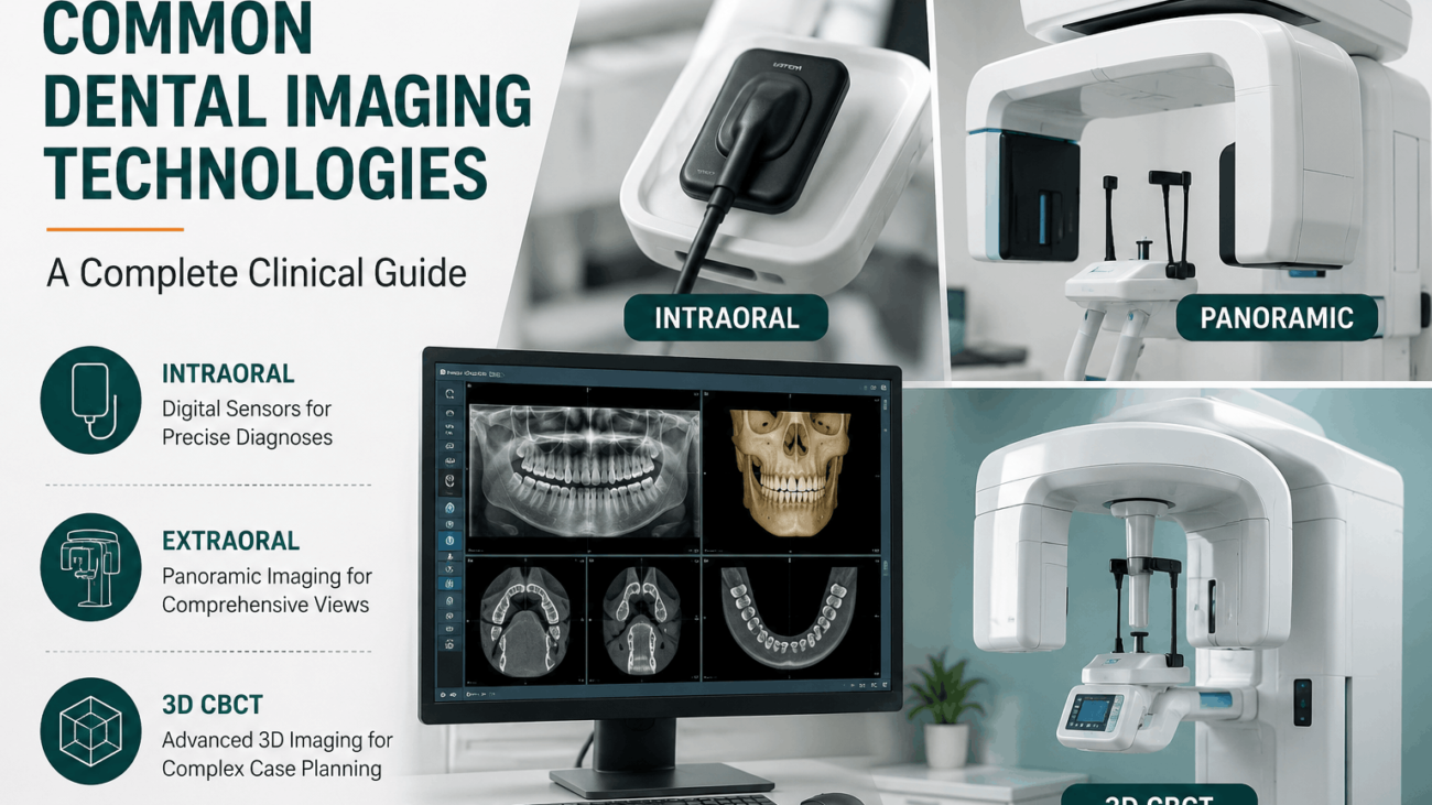
Common Dental Imaging Technologies
An engineering breakdown of modern diagnostic systems, tracking structural workflows from direct solid-state intraoral receptors up to 3D cone beam volumetric scans.
⏱️ 10-12 Min Read
✓ Comprehensive Clinical Hardware Guide
⚡ Summary of Modern Diagnostic Hardware
Modern dental practices rely on an integrated suite of **Common Dental Imaging Technologies** to identify pathologies hidden deep within soft tissues and bone matrices. These systems are broadly split into **intraoral technology**—such as high-efficiency, solid-state **CMOS digital sensors** and flexible **Phosphor Storage Plates (PSP)** placed inside the mouth for precise caries detection—and **extraoral technology**. Extraoral solutions feature **Digital Panoramic units** that map out the entire dental arch in a single sweep, alongside **Cone Beam Computed Tomography (CBCT)**, which renders true 3D volumetric images essential for complex root canal therapies, bone grafting procedures, and precision dental implant placements.
Table of Contents
→ 2. 2D Extraoral Diagnostic Hardware (Panoramic & Cephalometric)
→ 3. 3D Cone Beam Computed Tomography (CBCT Volumetric Imaging)
→ 4. Next-Gen Auxiliary Tools (Intraoral Cameras & Transillumination)
→ 5. Comprehensive Imaging Technology Comparison Matrix
→ 6. Frequently Asked Questions
1. Intraoral Imaging Receptors (CMOS vs. PSP Plates)
Intraoral modalities represent the most common foundational tier within the landscape of **Common Dental Imaging Technologies**. These systems involve placing a highly responsive receptor directly inside the patient’s oral cavity to capture localized structural views, such as interproximal bitewings, periapical root maps, or wide occlusal structures.
Solid-State Digital Sensors (CMOS)
Direct digital sensors built on **Complementary Metal-Oxide-Semiconductor (CMOS)** architecture stand as the gold standard for clinical speed. These rigid receptors are connected via a durable USB cord or a wireless transmitter block directly to your chairside computer monitor. The instant an exposure finishes, the image renders on-screen in less than two seconds, allowing the dental team to diagnose issues immediately without stepping away from the patient’s side.
Phosphor Storage Plates (PSP)
Phosphor storage systems utilize cordless, highly flexible plates that mirror the comfortable shape of traditional dental film. When an exposure occurs, the phosphor surface stores the raw energy data within its internal lattice. The plate is then placed inside a mechanical laser scanner that processes the digital file onto your screen before instantly clearing the plate for immediate sterilization and re-use.
2. 2D Extraoral Diagnostic Hardware
When a clinician needs a broader view that stretches past individual tooth roots, they shift toward extraoral imaging platforms. These systems keep the imaging receptors completely outside the patient’s mouth, utilizing synchronized mechanical sweeps to capture sweeping anatomical maps.
**Digital Panoramic Radiography** machines are a vital asset within this category. The x-ray source arm revolves smoothly around the patient’s head, generating a single, flattened 2D panoramic map that displays both jaw arches, the temporomandibular joints (TMJ), and the maxillary sinus cavities simultaneously. For specialized orthodontic practices, **Cephalometric attachments** are added to these systems to capture precise lateral skull bone proportions, which are critical for charting shifting orthodontic movements accurately.
3. 3D Cone Beam Computed Tomography (CBCT)
While 2D panoramic sweeps provide fantastic foundational summaries, flat images can occasionally camouflage deep structural overlaps. **Cone Beam Computed Tomography (CBCT)** resolves this limitation by providing true 3D volumetric rendering to modern dental operatories.
Unlike heavy medical CT scanners that rely on thin slice paths, a dental CBCT machine emits a highly focused, cone-shaped radiation beam. As the arm rotates around the patient, it captures hundreds of distinct high-resolution data slices in a single pass. The processing software compiles these data blocks into an interactive 3D model, allowing oral surgeons and endodontists to rotate bone maps, inspect hidden root canal branches, and plan dental implant paths with microscopic accuracy.
4. Next-Gen Auxiliary Imaging Systems
To round out a truly modern diagnostic workflow, clinics frequently add non-radiographic auxiliary technologies to their setups. **Intraoral Cameras** are small, pen-sized devices equipped with bright LED rings that take high-magnification, full-color photos of tooth surfaces. These systems serve as powerful patient education tools, allowing individuals to see structural fractures or margin leaks directly on a chairside screen.
Similarly, advanced **Digital Transillumination** devices pass intense, focused near-infrared light paths straight through tooth structures. Because dense, healthy enamel guides light differently than active decay networks, this technology allows clinicians to spot early interproximal caries without exposing the patient to any diagnostic radiation.
5. Comprehensive Imaging Technology Comparison Matrix
This analytical reference matrix compares the primary systems within modern dental imaging, helping you evaluate performance and clinical scope:
| Imaging Modality Technology | Image Format | Core Diagnostic Target Focus | Primary Workflow Advantage |
|---|---|---|---|
| CMOS Digital Sensors | High-Bit 2D Digital Map | Interproximal decay networks, fine root details, bone levels | **Instant chairside rendering** in under 2 seconds; zero processing lag. |
| Phosphor Storage Plates (PSP) | Flexible 2D Digital Scan | Caries detection, multi-size pediatric and adult views | Cordless, thin plate design provides **excellent patient comfort**. |
| Digital Panoramic Units | Wide 2D Comprehensive Map | Full dental arch alignment, TMJ paths, sinus boundaries | Captures the entire oral complex in **one single automated sweep**. |
| 3D CBCT Systems | 3D Volumetric Model | Implant path planning, bone volume, root canal paths | Delivers comprehensive **three-dimensional architectural precision**. |
| Intraoral Cameras | Full-Color High-Mag Photo | Surface fractures, soft tissue marks, old crown margins | **Exceptional patient education tool** for visual verification. |
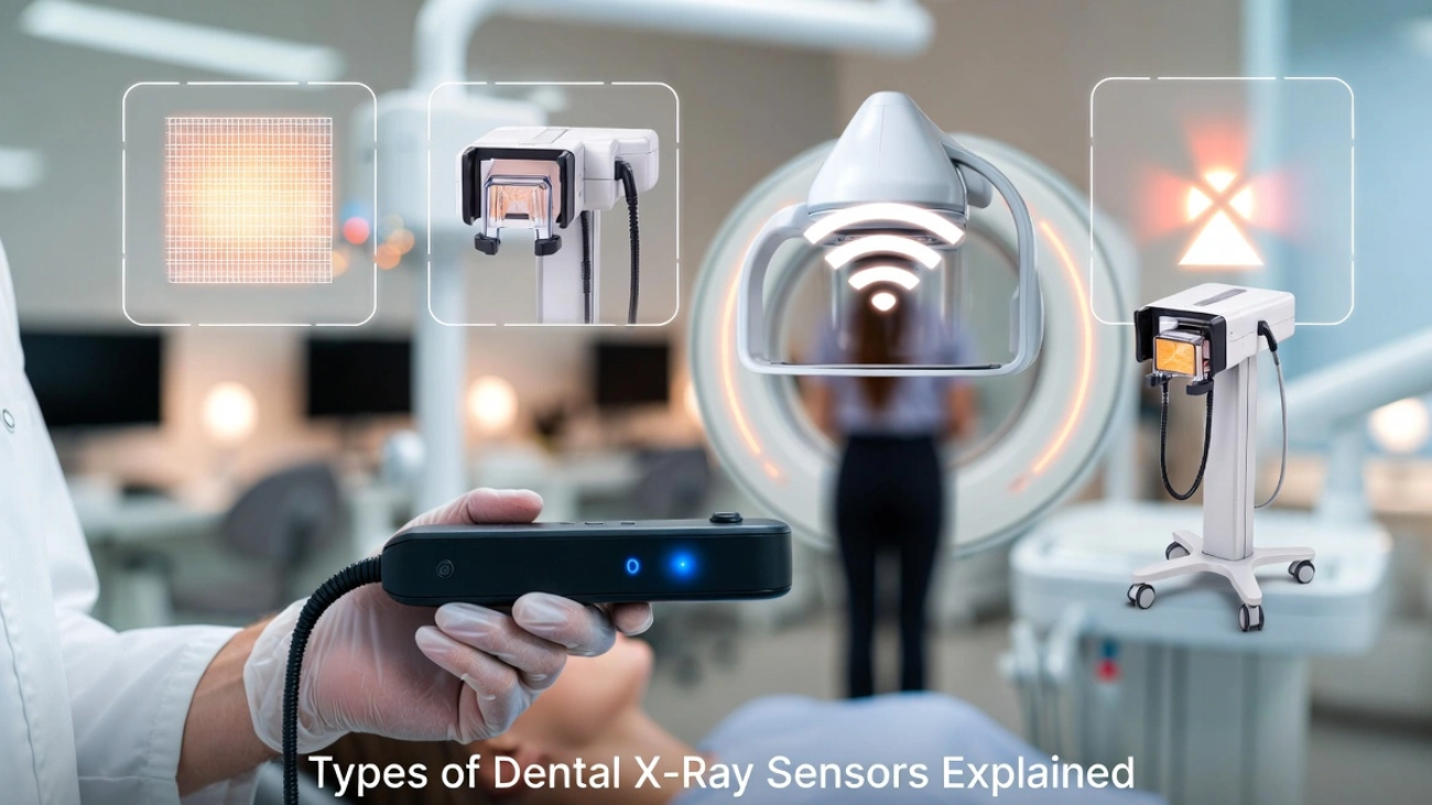
🔬 Go Deeper Into Sensor Engineering: To truly understand the internal silicon structures and scintillator physics that power these high-speed intraoral systems, explore our detailed guide explaining exactly how dental x-ray sensors work to master direct-conversion engineering and pixel architectures.
6. Frequently Asked Questions
What is the practical difference between choosing a CMOS sensor over a PSP system?
CMOS sensors provide direct data processing straight to your screen in under two seconds, optimizing fast-paced operations. PSP systems require a manual scanning step, but they offer greater plate flexibility, which helps when accommodating patients with shallow palates or sensitive anatomy.
Why are 3D CBCT scans preferred over 2D periapical options for dental implant placements?
Standard 2D images cannot show the true physical thickness or width of the jawbone, which introduces risk when plotting implant posts. A 3D CBCT scan provides cross-sectional views that allow doctors to precisely evaluate bone volume and avoid sensitive nerve paths.
Do non-radiographic transillumination devices entirely replace standard x-rays?
No. While transillumination tools excel at spotting early interproximal enamel fractures without radiation, they cannot penetrate deep bone layers or trace underlying root infections, meaning standard digital sensors remain a clinical necessity.
Modernize Your Clinic With Elite Imaging Systems
Equip your practice with state-of-the-art diagnostic power. Discover top-tier digital CMOS sensors, smart PSP laser scanners, and advanced 3D CBCT configurations at SwatDental.