Digital vs Traditional Dental X-Rays
A deep technical evaluation comparing solid-state digital sensors against legacy analog film envelopes across diagnostic resolution, patient dosing safety, and operatory costs.
⏱️ 10-12 Min Read
✓ Technical Safety Compliant
⚡ Core Technology Paradigm Shift
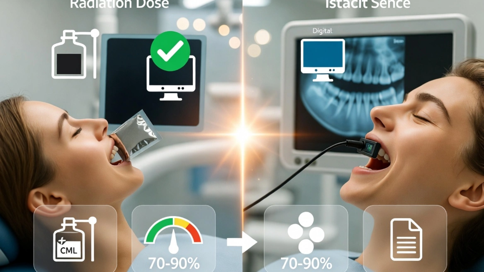
The central difference when breaking down **Digital vs Traditional Dental X-Rays** lies in the **capture mechanism and processing time**. Traditional X-rays rely on slow, analog film packets that require chemical development in a darkroom using toxic fixing agents, imposing a 5-to-8 minute operational delay. Conversely, digital radiography leverages sensitive solid-state silicon sensors (**CMOS/CCD**) or reusable phosphor sheets (**PSP**) that immediately translate radiation into high-bit pixel maps on a monitor. This shift to digital reduces patient **radiation exposure by up to 70% to 90%**, eliminates hazardous chemical waste, and unlocks advanced software manipulation filters for vastly sharper diagnostic precision.
Table of Contents
1. Radiation Dosing & Patient Safety Metrics
When patients evaluate the topic of **Digital vs Traditional Dental X-Rays**, radiation exposure is almost always their primary concern. Traditional film radiography uses silver halide grains suspended inside a plastic sheet. Because these physical film grains require a relatively massive volume of photon strikes to trigger a dark chemical reaction, the patient must be exposed to longer radiation bursts from the tubehead.
Modern digital sensors alter this safety dynamic entirely. High-tier solid-state **CMOS receptors** boast exceptionally high **Quantum Efficiency (QE)**. Because the silicon pixels are highly responsive to photon packets, they require significantly less radiation to output a perfectly saturated image. Transitioning to digital channels slashes patient exposure settings by **70% to 90%** depending on whether you are shifting from D-speed or faster F-speed film, aligning perfectly with the strict **ALARA (As Low As Reasonably Achievable)** clinical safety protocols.
2. Darkroom Chemicals vs. Immediate Screen Rendering
The day-to-day operatory workflow represents another massive point of divergence when comparing **Digital vs Traditional Dental X-Rays**. The difference in image rendering mechanics directly dictates how many patients your team can smoothly care for in a shift.
Traditional Film Processing Cycles
Taking an analog film X-ray requires a tedious sequence of manual mechanical steps:
- Place the plastic film envelope in the patient’s mouth.
- Expose the packet, then walk it down the hall to a light-locked darkroom or counter-top processor box.
- Unwrap the packet carefully, feed the film sheet into developer fluid tanks, rinse it in water, slide it into fixing chemicals, and wait for a full drying cycle.
This entire process introduces a **5-to-8 minute delay**. If the assistant accidentally misaligned the tubehead angle slightly, the error isn’t discovered until the dried film is ready, forcing a frustrating re-shoot sequence that breaks your schedule flow.
Digital Immediate Acquisition Channels
Direct digital sensors compress this entire workflow into a matter of moments. The solid-state chip rests securely inside a hygienic protective sleeve and captures the image data instantly. Within **less than two seconds**, the completed scan flashes onto the treatment monitor right next to the patient’s chair.
The clinician remains comfortably at the bedside the entire time. If an exposure adjustment or angle modification is needed, it can be executed instantly, creating a vastly smoother experience for both the assistant and the patient.
3. Spatial Resolution & Software Image Enhancement
At first glance under perfect laboratory setups, traditional high-quality analog film can display strong spatial resolution marks. However, in the real-world daily practice of analyzing **Digital vs Traditional Dental X-Rays**, digital channels possess a decisive diagnostic advantage thanks to software manipulation.
When a traditional film is processed, the resulting image is static. If the exposure turns out slightly too dark or too light, you must rely on a standard desk lightbox and a magnifying glass to squint at the small 2-inch plastic sheet, searching for subtle anomalies.
Digital imaging software transforms this passive review into an active diagnostic tool. Once the digital pixel map lands on your monitor, you can immediately **zoom in, adjust contrast levels, and invert grayscale ranges**. Specialized filters can sharpen edge boundaries to spotlight initial interproximal decay networks, trace tricky bone loss lines, or inspect apical root paths with incredible precision.
4. Environmental Impact & Practice Economics
Looking past direct clinical variables reveals a stark contrast in the environmental footprints and backend business costs of **Digital vs Traditional Dental X-Rays**. Traditional systems generate a continuous stream of material waste and chemical overhead. Practices must constantly purchase film packets, lead backing sheets, and fresh gallons of developer and fixer fluids.
Disposing of these depleted chemicals is heavily regulated because they carry hazardous heavy metal ions. This requires specialized waste collection contracts that add ongoing financial overhead to your office ledger.
While switching to a digital workflow involves an initial hardware investment in solid-state sensors, it eliminates these recurring material costs entirely. Your practice completely wipes away darkroom chemical maintenance, plastic film packet garbage, and physical chart filing cabinets. Digital scans are saved straight to local servers or secure cloud platforms, making records instantly shareable with insurance companies or specialists via encrypted links.
5. Side-by-Side Performance Comparison Matrix
This comprehensive table summarizes the core differences between **Digital vs Traditional Dental X-Rays** to help guide your practice conversion plans:
| Performance Metric | Traditional Analog Film Systems | Modern Digital Sensor Platforms |
|---|---|---|
| Patient Radiation Exposure | **Higher:** Requires extended emission timing to activate film grain matrices. | **Significantly Lower:** Slashed by **70% to 90%** due to high QE silicon logic. |
| Image Acquisition Speed | **Delayed:** Requires **5 to 8 minutes** for complete chemical tank cycling. | **Instantaneous:** Renders on operatory screens in **under 2 seconds**. |
| Diagnostic Viewing Options | **Static:** Limited to viewing small physical sheets over a desk lightbox. | **Dynamic:** Full-screen zoom, live contrast tweaks, and sharp edge filters. |
| Chemical & Waste Footprint | **High:** Constant disposal of heavy lead sheets and toxic chemical fluid tanks. | **Zero:** Entirely paperless and chemical-free electronic data path files. |
| Record Storage & Sharing | **Physical:** Stored in manual chart jackets; requires physical mail for transfers. | **Digital:** Instantly integrated into dental software; simple encrypted link sending. |
| Recurring Material Overhead | **High:** Ongoing costs for film packets, developer fluids, and toxic waste disposal. | **Minimal:** Bound entirely to initial sensor asset purchases and barrier sleeves. |
🔗 Explore the Semiconductor Architecture: If you want to understand the exact internal layer stack configurations that enable this massive radiation reduction, read our detailed technical entry explaining how dental x-ray sensors work to master scintillator physics and chip layouts.
6. Frequently Asked Questions
Can I use my existing wall-mounted tubehead machine if I switch to digital sensors?
Yes, almost all traditional X-ray tubeheads work perfectly with digital sensors. You simply need to adjust the timer setting down to match the shorter exposure intervals required by digital silicon chips.
Is a digital dental sensor uncomfortable for patients compared to soft film?
Rigid solid-state digital sensors are thicker than traditional film packets. However, modern sensor brands counter this by designing smooth, heavily rounded corners and offering multiple sizes to match different patient anatomies cleanly.
How are digital dental X-ray files protected under modern data privacy rules?
Digital radiography files are stored directly inside your practice management software, which secures patient data through advanced local database encryption, user password paths, and encrypted cloud backup networks.
Ready to Transition to Modern Digital Diagnostics?
Stop spending money on recurring film packets and toxic chemical lines. Explore premium, high-efficiency digital imaging configurations at SwatDental to modernize your clinical workflow today.
Add a Comment