Are Dental X-Rays Safe?
Demystifying diagnostic radiation exposure using concrete scientific benchmarks, millisievert data, and the protective engineering inside modern imaging hardware.
⏱️ 8-10 Min Read
✓ Verified by Radiology Standards
⚡ The Definitive Clinical Answer
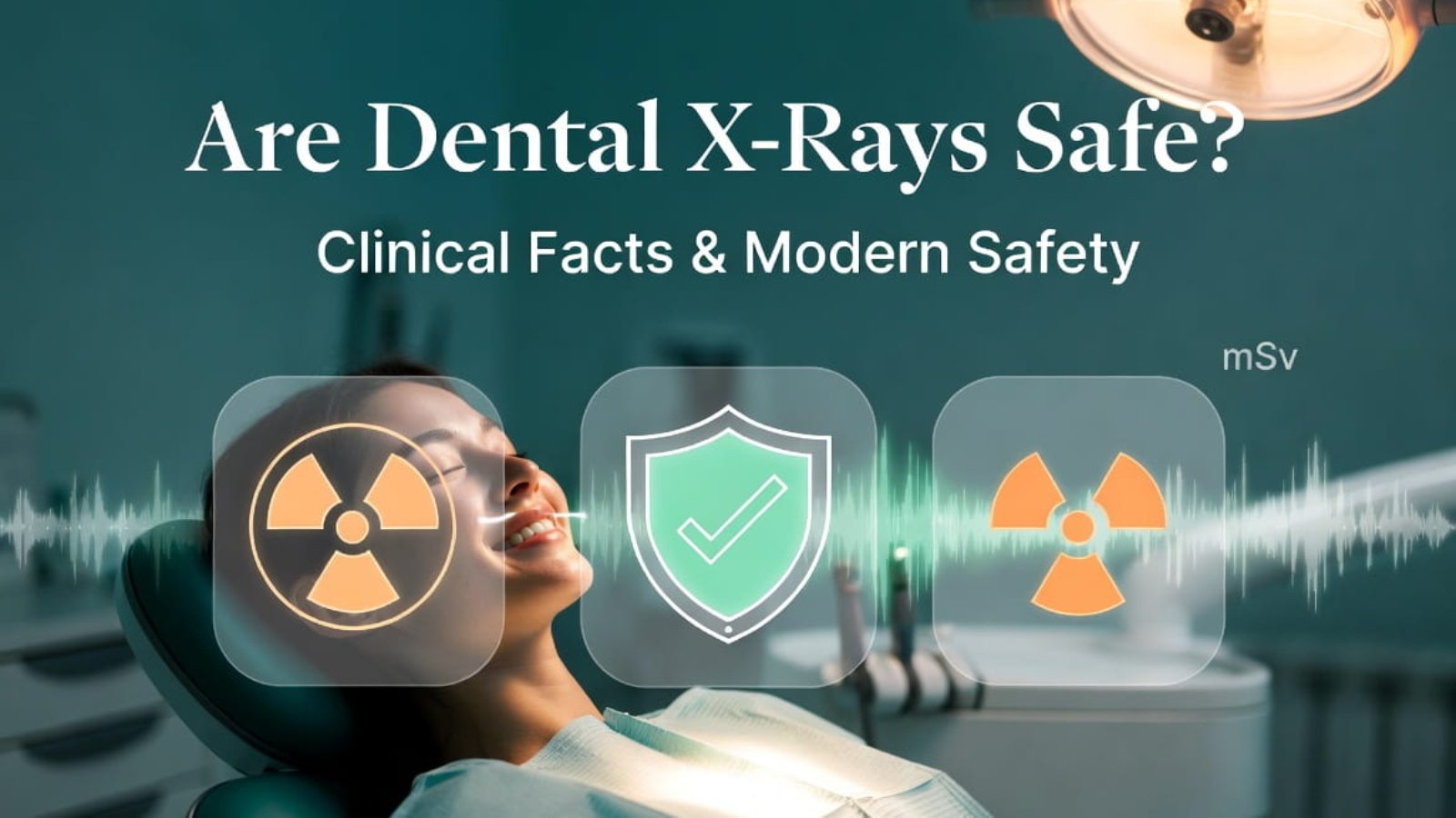
Yes, **modern dental X-rays are exceptionally safe**. When asking **Are Dental X-Rays Safe?**, it helps to look at the numbers: a standard set of digital intraoral bitewings exposes a patient to roughly **0.005 millisieverts (mSv) of radiation**. This tiny amount is equivalent to what you naturally absorb from the sun and soil during just **less than one single day of normal life on Earth**. Thanks to advanced solid-state **CMOS digital sensors**, lead-lined thyroid collars, and tightly focused radiation beams, the medical risk is incredibly low, while the diagnostic benefit of identifying underlying bone loss, root infections, and deep decay is safely maximized.
Table of Contents
→ 2. Real-World Radiation Dosing Benchmark Matrix
→ 3. How Digital Technology Redefined Patient Safety
→ 4. Triple-Layer Protection Protocols (ALARA, Aprons, Collars)
→ 5. Diagnosing vs. Guessing: The Hidden Hazard of Refusal
→ 6. Frequently Asked Questions
1. Putting Radiation into Perspective: The mSv Metric
The word “radiation” often triggers immediate anxiety, but the key to addressing whether **Are Dental X-Rays Safe?** is understanding the concept of dosage scale. In medical physics, effective radiation absorption is calculated using a metric called **Millisieverts (mSv)**.
Human beings are exposed to natural background radiation every second of their lives. Radioactive isotopes in the earth’s rocky layers, cosmic rays hitting our atmosphere from deep space, and even natural potassium traces in food items create a steady baseline exposure. On average, a typical person absorbs roughly **3.0 mSv of background radiation every year** simply by existing on the planet. When contrasted against this continuous background baseline, the electrical emission from a focused dental image is minor.
2. Real-World Radiation Dosing Benchmark Matrix
To truly understand the question—**Are Dental X-Rays Safe?**—it helps to see how dental diagnostics compare to other everyday sources of radiation exposure:
| Exposure Event Source | Approximate Radiation Dose (mSv) | Equivalent Natural Background Time Baseline |
|---|---|---|
| Digital Intraoral Bitewings (4 Images) | **0.005 mSv** | Less than 1 Single Day of normal living |
| Digital Panoramic Extraoral Scan | **0.010 to 0.015 mSv** | Roughly 1 to 2 Days of normal living |
| Cross-Country Airline Flight (NY to LA) | **0.035 mSv** | About 4 Days of elevated cosmic ray exposure |
| Annual Natural Background Intake (Global Average) | **3.000 mSv** | 365 Days of standard environmental background |
| Standard Medical Chest X-Ray | **0.100 mSv** | About 10 Days of normal living |
| Medical Lumbar Spine Series | **1.500 mSv** | Roughly 6 Months of normal living |
| Full Medical Abdominal CT Scan | **10.000 mSv** | Roughly 3 Years of accumulated natural baseline exposure |
3. How Digital Technology Redefined Patient Safety
The conversation around safety has completely shifted over the last decade thanks to the decline of traditional analog film packets. Old-school film setups relied on silver halide crystals, which required a longer, heavier burst of radiation to register an image on the plastic sheet.
Today’s **modern digital CMOS sensors** use highly advanced active pixel technology. These solid-state silicon chips catch incoming photons and convert them into clear digital images with incredible efficiency. This technological leap allows modern practices to cut down patient radiation exposure by **70% to 90%** compared to old film methods, making an already low-risk procedure practically microscopic.
4. Triple-Layer Protection Protocols
In addition to utilizing fast digital sensors, dental teams use a multi-layered safety strategy to ensure patient exposure stays as close to zero as possible:
The ALARA Principle
Standing for **As Low As Reasonably Achievable**, this is the guiding rule for clinical radiography. It means a doctor will never take an image just because a calendar says so. Every single scan must have an explicit diagnostic reason based on a physical check of your teeth.
Leaded Aprons & Thyroid Collars
Even though modern X-ray beams are incredibly focused, protective aprons lined with lead or composite metals block any secondary scatter radiation. A built-in thyroid collar fits securely around the neck, completely shielding sensitive hormone glands from unnecessary exposure.
Precision Beam Collimation
Modern X-ray tubeheads use long, lead-lined cones called collimators. Instead of letting radiation spread out in a wide circle, a rectangular collimator narrows the beam down to the exact size and shape of the sensor, protecting the surrounding facial tissues perfectly.
5. Diagnosing vs. Guessing: The Hidden Hazard of Refusal
When considering **Are Dental X-Rays Safe?**, it’s crucial to look at the other side of the equation: the very real danger of refusing necessary diagnostic images. A standard visual look with a dental mirror can only see the outer surfaces of your enamel.
More than **50% of an adult’s tooth structure** hidden beneath the gum line remains completely invisible to the naked eye. Without digital imaging, serious issues can develop undetected, including:
- Hidden decay tunneling between tight tooth contacts.
- Asymptomatic infections brewing deep around root tips or bone structures.
- Silent jawbone loss caused by progressive periodontal disease.
- Cysts, abscesses, or impacted teeth threatening neighboring roots.
By the time these hidden anomalies cause physical pain, the damage is often extensive, requiring complex treatments like root canals or extractions. Catching these issues early with low-dose digital imaging is the safest way to preserve your long-term health.
🔬 Go Deeper Into the Technology: Want to see exactly how these low-dose systems turn minor photon signals into crisp images? Read our comprehensive technical guide explaining how dental x-ray sensors work to learn about internal scintillator screens and direct conversion engineering.
6. Frequently Asked Questions
Are dental X-rays safe for young children?
Yes, they are completely safe. Children’s developing mouth structures actually make them prime candidates for early monitoring. Modern digital systems feature customized pediatric sizing modes that reduce exposure to a tiny fraction of adult settings.
Can dental radiography increase my long-term medical risks?
The risk of developing complications from a modern digital dental scan is practically zero. You absorb far more radiation from everyday cosmic rays during a routine commercial flight than you ever will from a localized bitewing set.
Why does the assistant leave the room if dental X-rays are safe?
While a single X-ray dose is minor for a patient, dental assistants take dozens of scans every single day. Stepping behind a lead-shielded wall protects the clinical team from accumulating repetitive exposure over years of practice.
Upgrade to Ultra-Low Radiation Standards
Give your patients absolute peace of mind. Equip your practice with the industry’s safest high-efficiency digital CMOS sensors and portable, heavily shielded X-ray tech from SwatDental.
Add a Comment